Comparison of mechanical thrombectomy techniques in an in vitro stroke
model: How to obtain a first pass recanalization?
Gascou Gregorya, Cagnazzo Federicoa, Pierre-Henri Lefevrea, Dargazanli Cyrila,
Costalat Vincenta, Omer Faruk Ekerb
Department of neuroradiology, Hopital Gui de Chauliac, Montpellier, France
Department of neuroradiology, Hopital Pierre Wertheimer, Bron, France
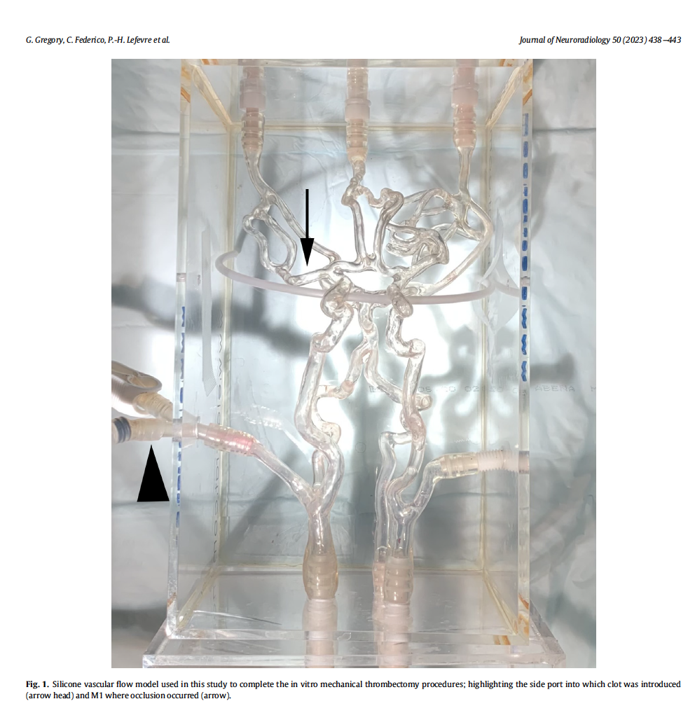
Silicone vascular in vitro model : 
Simulated in vitro MT procedures were performed using a realistic soft 3-D silicone model (Elastrat Sarl, Geneva, Switzerland) of the human cerebral vasculature with complete circle of Willis
Download PDFTo Balloon or Not to Balloon? The Effects of an Intra-Aortic Balloon-Pump on Coronary Artery Flow during Extracorporeal
Circulation Simulating Normal and Low Cardiac Output Syndromes.
Philippe Reymond, Karim Bendjelid, Raphaël Giraud, Gérald Richard, Nicolas Murith, Mustafa Cikirikcioglu and Christoph Huber.
Download PDFMethodologies to assess blood flow in cerebral aneurysms : current state of research and perspectives. Study developed with a Elastrat intracranial aneurysm model.
Download PDFEffect of Flow Diverter Porosity on Intraaneurysmal Blood Flow. Study developed with a Elastrat intracranial aneurysm model.
Download PDFComparison of the Effectiveness of Three Methods of Recanalization in a Model of the Middle Cerebral Artery: Thrombus Aspiration via a 4F Catheter, Thrombus Aspiration via the GP
Download PDFArtificial models of cerebral aneurysms for medical training and testing of medical devices were con- structed from corrosion casts of the main cerebral arteries of a human specimen. Three aneurysms with a variety of shapes were simulated at typical locations. Rigid and soft models were made of silicone using the ``lost wax'' technique. The transparent silicone models were anatomically accurate and reproducible copies of human vascular casts. These models could be connected in a closed circuit that used an electric pump to simulate pulsatile flow.
Endovascular procedures and surgical clip application were performed under fluoroscopic or direct visual control. Surgical clipping, endoluminal coil manipulation, and consecutive hemodynamic changes were visualized by digital subtraction angio- graphy and direct observation. The model provides trainee surgeons with an understanding of clinical conditions. New medical devices, such as platinum coils, would be experimentally implanted in the model under stable conditions. These anatomically accurate and reproducible models of cerebral vas- culature and aneurysms are valuable for medical testing, training, and research.(® All rights reserved).
Download PDFComputed flow dynamics simulation of a patient on a specific Head and Neck custom made model developed by Elastrat after 3D reconstruction on a flat detector angiograohic system.
Velocity stream lines and wall shear stress provided by this computation could give a significant information for the clinician about intra aneurysmal flow patterns and stimulation of aneurysm wall.
Embolus trajectory through a physical replica of the major cerebral arteries. Elastrat used model H N-S-A-010.
(From "Stroke", the Journal Of The American Heart Association – All rights reserved).
Laser Doppler Velocimetry and PC-MRA validated in an anatomical cerebral artery model.
Download PDFExperimental study of aortic flow in the ascending aorta via Particle Tracking Velocimetry
Download PDFValidation in an Anatomically Accurate Cerebral Artery Aneurysm Model With Steady Flow
Download PDFStudy of in-vitro models of human carotid atheromatous disease
Download PDFSilicon models as basic training and research aid in endovascular neurointervention.
Download PDFGeometry and renal flow in chimney-Endovascular Aneurysm Repair and chimney-Endovascular Aneurysm Sealing. June 2017
Download PDF
Copyright © Elastrat. All right reserved - LEGAL NOTICE - PRIVACY POLICY
Elastrat Sàrl is a worldwide leader in the development, realization and distribution of human anatomical vascular phantoms.
Our Swiss made transparent soft or rigid silicone phantoms reproduce the anatomical vasculature with its pathologies like aneurysms or stenoses and can be trained through navigation with stents, catheters and coils. They provide a realistic environment for the simulation of endovascular procedures, pre-surgery training, studies and teaching purposes for interventionists, such as interventional cardiologists (neuroradiologists, etc.), interventional radiology-cardiologists, and interventional angiologists.
The models promote safety through education and professional competence.

